How the Eye Works
Chicago, Illinois
Although vision can be described simply and easily as being like taking a photo with a traditional camera, the eye’s structures are tiny and delicate, and their interactions are complex. Protected by the optic rim of the skull, the eye consists of a mostly fluid-filled sphere that uses its structures to focus light and convert it to electrical energy for the brain to interpret. We are all aware of the white of the eye and the color of a person’s eye, but many important structures are at work beneath the surface.
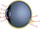
The Cornea is the transparent membrane that curves over the eye’s surface. The "twinkle of the eye" is seen in the surface of the cornea. It bends, or refracts, light to focus it on the retina at the back of the eye. It is the cornea that determines the acuity of one’s eyesight according to its shape or curvature accounting for nearsightedness, farsightedness and astigmatism. The cornea connects to the sclera and is part of the eyeball wall.
The Iris is a muscular ring behind the cornea and is the colored part. It can be blue, green, grey brown or some other combinations of color. It controls the size of the pupil at its center, regulating the amount of light that enters the eye. The pupil is not an actual structure, just an aperture (or hole) in the iris. The pupil looks black in normal healthy eyes.
The Sclera is the white of the eye and is part of the eyeball wall which continues around to the retina at the back. It connects to the cornea. It is the structure of the eye that give it strength and maintains the ball-like shape.
The Lens is located behind the iris in a fluid-filled chamber called the posterior chamber. The lens further bends light traveling in from the cornea to make it focus on the retina. Tiny muscles around the lens control its curvature, making it steeper for near vision and flatter for distance vision. In a 20/20 eye, the lens and cornea work well together and focus all incoming light on the retina, providing clear vision at all distances. It is the lens that becomes cloudy with old age and this is called a cataract.
The Retina is the equivalent of camera film at the back of the eye. It forms the back wall of a fluid-filled chamber called the vitreous chamber, which makes up most of the eye’s entire mass. Retinal cells are light-sensitive and of two types: (a) cones, mostly clustered near the center of the retina, which give us our direct vision in bright light and our color perception; and (b) rods, mostly around the retinal periphery, which give us grayscale vision in dim light.
In the center of the retina is an area called the macula, where cones are most densely clustered; and nearby is the eye’s blind spot, an area of no light-sensitive cells, where the optic nerve fibers converge to leave the eye.
The Optic Nerve consists of millions of nerve fibers, each serving one or more retinal cells. They pick up the electrical impulses converted by the cells from light energy and run across the retina, converging to form the optic nerve which leaves the retina in a nerve sheath. It travels to the brain’s vision center like a telephone cable where the electrical impulses are interpreted as images.
The fluid in the anterior chamber is called aqueous humor. The lens and cornea have no blood supply, as blood vessels would block light and impair vision. Aqueous fluid bathes the lens through small openings and provides necessary nutrients to the lens and cornea. The fluid behind the lens is called vitreous humor, and although mostly water, has a gel like consistency which helps maintain the shape of the eye.
The eye also contains glands for tear production and both sensory and motor nerves that register pressure, pain etc. and allow eye movement and focus.
The eye is a complex organ, requiring knowledgeable and experienced professional treatment. The dedicated professional team at Doctors for Visual Freedom Laser Center is trained and ready to diagnose and treat vision problems and diseases of the eye. We offer LASIK surgery to correct nearsightedness, farsightedness and astigmatism.
Learn more about general eye care and eye care facts and myths.
If you live in the Chicago, Arlington Heights or Downtown Chicago metropolitan area, please contact our experienced team at Doctors for Visual Freedom Laser Center today.
Two Locations:
Doctors for Visual Freedom Laser Center
875 North Michigan Avenue, Suite 1550
Chicago, IL 60611 | Doctors for Visual Freedom Laser Center
2010 S Arlington Heights Rd, Suite 121
Arlington Heights, IL 60005 |